
Foot & Ankle X-Rays
Foot and ankle x-rays are a common diagnostic tool used by podiatrists and other medical professionals to visualize the bones and tissues within the foot and ankle. X-rays use a small amount of radiation to create an image of the inside of the body, which can help to identify any issues or abnormalities.
This article will provide a comprehensive guide to foot and ankle x-rays, including when they are typically ordered, what to expect during the procedure, and how the results are interpreted. We will also discuss this diagnostic test's potential risks and limitations and alternative techniques that may be used to diagnose foot and ankle issues.
What are Foot and Ankle X-Rays?
Foot and ankle x-rays are a medical imaging test that uses a small amount of radiation to create detailed images of the bones and tissues within the foot and ankle. These images can help medical professionals diagnose various conditions, including fractures, dislocations, and other injuries.
How X-Rays Work
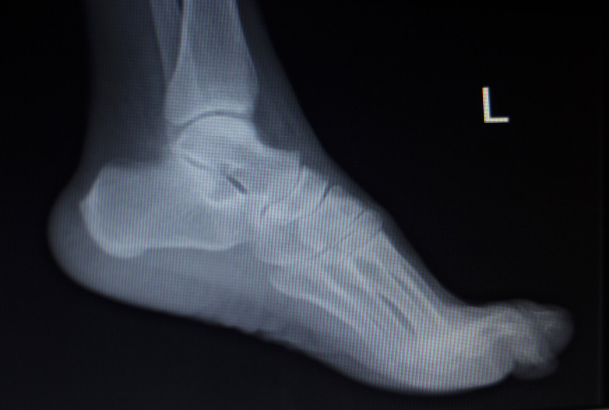
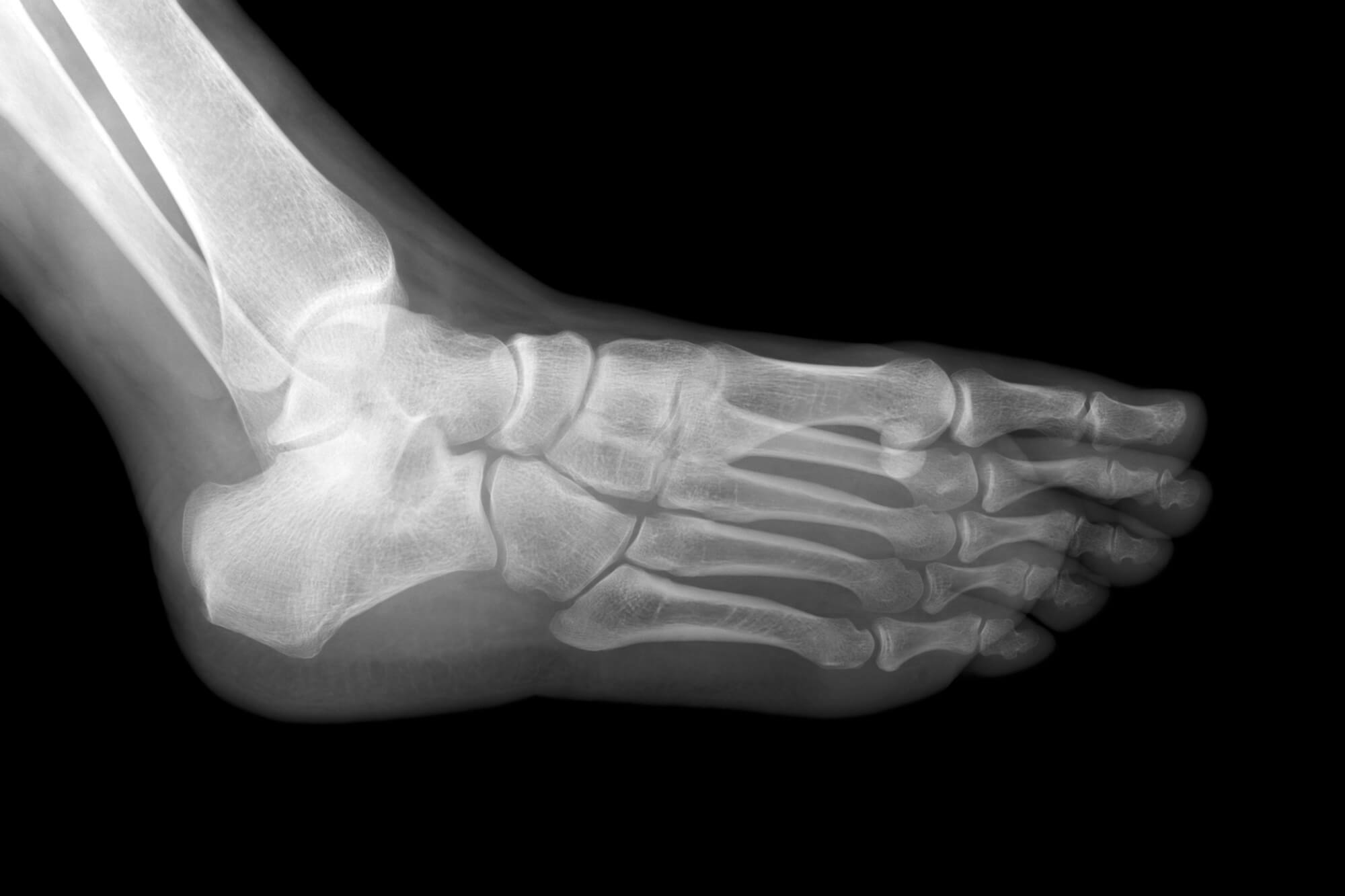
X-rays work by using a special machine that produces a beam of radiation, which passes through the body and is absorbed by different tissues at different rates. Dense tissues, such as bones, absorb more radiation and appear white in the resulting image. Softer tissues, such as muscles and organs, absorb less radiation and appear gray in the image. The resulting image is a radiograph, which can be used to visualize the anatomy of the foot and ankle in great detail.
Benefits of Foot and Ankle X-Rays
Foot and ankle x-rays have several benefits, including:
- They are quick and easy to perform, with the entire procedure taking just a few minutes.
- They are relatively painless and do not require any injections or incisions.
- They provide detailed images of the bones and tissues within the foot and ankle, which can help medical professionals accurately diagnose a wide range of conditions.
- They are widely available and can be performed in most hospitals and clinics.
When are Foot and Ankle X-Rays Ordered?
Foot and ankle x-rays are typically ordered by a podiatrist or other medical professional when there is a need to visualize the bones and tissues within the foot and ankle. Some common reasons for ordering foot and ankle x-rays include the following:
Suspected Fractures or Dislocations
If a patient has suffered an injury to the foot or ankle, such as a fall or sports-related injury, a foot and ankle x-ray may be ordered to check for any fractures or dislocations. These conditions can be difficult to diagnose based on physical examination alone, and an X-ray can help to confirm the diagnosis and determine the appropriate treatment.
Foot or Ankle Pain with Unknown Cause
Sometimes, a patient may experience foot or ankle pain with an unknown cause. An X-ray can help to identify any underlying issues, such as a fracture or osteoarthritis, that may be causing the pain.
Pre-Surgery Planning
Foot and ankle x-rays may also be ordered as part of planning certain surgeries, such as joint replacement surgery. The X-rays can help the surgeon to understand the anatomy of the foot and ankle and plan the surgery accordingly.
What to Expect During the X-Ray Procedure
If a foot and ankle x-ray is ordered, the patient can expect the following during the procedure:
Preparation for the X-Ray
Before the X-ray procedure, the patient may be asked to remove any jewelry or other metal objects that may interfere with the X-ray image. The patient may also be asked to wear a hospital gown or other protective clothing.
The X-Ray Procedure
During the X-ray procedure, the patient will be asked to stand or sit in a certain position, depending on the area of the foot or ankle being imaged. The X-ray machine will be positioned a few feet away from the patient, and a beam of radiation will be directed towards the foot or ankle. The patient will be asked to remain still during the X-ray, which typically takes just a few seconds.
After the X-Ray
After the X-ray is complete, the patient can return to normal activities. There is usually no downtime or recovery after a foot and ankle x-ray.
Interpreting the Results of Foot and Ankle X-Rays
After the foot and ankle x-ray has been completed, the resulting images will be reviewed by a radiologist or a medical imaging specialist. The radiologist will look for any abnormalities or issues within the bones and tissues of the foot and ankle and report their findings to the ordering physician.

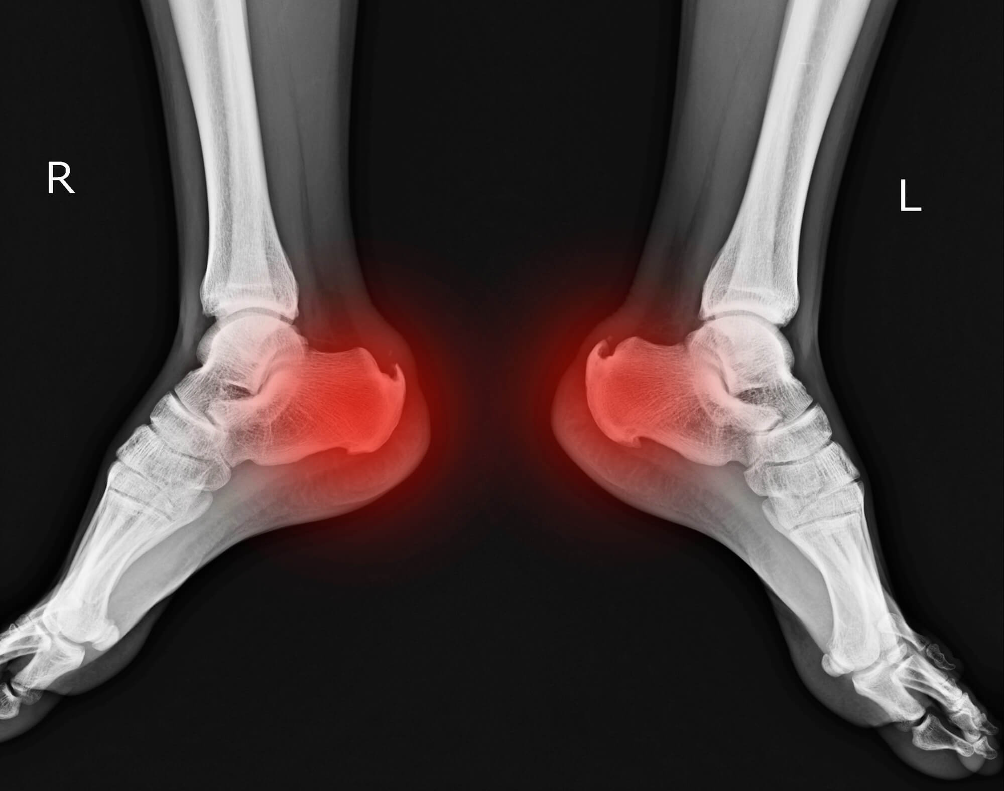
Common Findings on Foot and Ankle X-Rays
There are many potential findings on a foot and ankle x-ray, including:
- Fractures: A fracture is a bone break that can occur due to injury or overuse. Fractures can be seen on an X-ray as a line or gap within the bone.
- Dislocations: A dislocation is when a bone is displaced from its normal position, often due to injury. Dislocations can be seen on an X-ray as a misalignment of the bones.
- Osteoarthritis: Osteoarthritis is a type of degenerative joint disease that can cause the cartilage within a joint to break down. Osteoarthritis can be seen as a loss of joint space or bone spurs on an X-ray.
- Other abnormalities: Other abnormalities may be seen on a foot and ankle x-ray, such as cysts or tumors.

Follow-Up Care Based on X-Ray Results
Based on foot and ankle x-ray findings, the ordering physician may recommend further testing or treatment. This may include:
- Physical therapy: If a patient has suffered an injury or has a condition such as osteoarthritis, physical therapy may be recommended to help improve strength and mobility.
- Medication: The physician may prescribe medication to help reduce inflammation or pain.
- Surgery: In some cases, surgery may be necessary to repair a fracture or correct a deformity.
Risks and Limitations of Foot and Ankle X-Rays
While foot and ankle x-rays are generally safe and have few risks, there are some potential risks and limitations to be aware of:
Risks of X-Ray Exposure
Like all medical procedures that use radiation, there is a small risk of exposure to X-rays. However, the radiation used in a foot and ankle x-ray is generally very low and is considered safe for most patients.
Limitations of X-Ray Imaging
There are also some limitations to using X-ray imaging for the foot and ankle. For example:
- X-rays do not provide a detailed image of soft tissues, such as muscles and tendons.
- In some cases, the bones may overlap or be obscured by other structures, making it difficult to visualize certain areas.
- X-rays are not always effective at identifying issues within the soft tissues of the foot and ankle, such as ligament or tendon injuries.
Alternative Diagnostic Techniques
In addition to foot and ankle x-rays, several other diagnostic techniques may be used to evaluate foot and ankle issues, including:
- Magnetic Resonance Imaging (MRI): MRI uses a strong magnetic field and radio waves to create detailed images of the body's soft tissues. MRI is often used to evaluate injuries to the ligaments, tendons, and other soft tissues of the foot and ankle.
- Computed Tomography (CT) Scan: A CT scan uses X-rays and a computer to create detailed cross-sectional images of the body. CT scans often evaluate fractures and other foot and ankle bone injuries.
- Ultrasound: Ultrasound uses high-frequency sound waves to create images of the body's tissues. Ultrasound is often used to evaluate issues within the soft tissues of the foot and ankle, such as tendonitis or plantar fasciitis.
Conclusion
Foot and ankle x-rays are a quick, easy, and non-invasive way to visualize the bones and tissues within the foot and ankle. They can diagnose various conditions, including fractures, dislocations, and osteoarthritis. While there are potential risks and limitations, foot and ankle x-rays are generally considered safe. They are an important tool in diagnosing and treating foot and ankle issues.
In addition to foot and ankle x-rays, several other diagnostic techniques may be used to evaluate foot and ankle issues, including MRI, CT scans, and ultrasound. Overall, foot and ankle x-rays can help patients and their healthcare providers to identify and correct any issues within the foot and ankle, leading to improved health and mobility.