
Understanding X-rays for Heel Pain
Heel pain can be a debilitating condition that affects many people at some point in their lives. It can be caused by a variety of conditions, including plantar fasciitis, heel spurs, and fractures. X-rays are a common diagnostic tool used by podiatrists to help determine the cause of heel pain and develop a treatment plan.
In this article, we will explore what X-rays are, why they are used for heel pain, what to expect during an X-ray, how to understand the results, and treatment options based on the findings.
What Are X-Rays and How Do They Work?
What Are X-Rays?
X-rays are a form of electromagnetic radiation with wavelengths shorter than visible light. They are used to create images of the inside of the body, including bones, organs, and tissues. X-rays are non-invasive and painless, and they do not produce any lasting effects on the body.
How X-Rays Work
X-rays work by passing through the body and being absorbed by different tissues at different rates. Dense tissues such as bones absorb more X-rays than softer tissues such as muscles and organs. The X-rays that pass through the body are detected by a special film or digital detector, which creates an image of the inside of the body. The different densities of tissues appear as different shades of gray on the X-ray image, allowing the healthcare provider to see the structure and condition of the bones, organs, and tissues.
Why Are X-Rays Used for Heel Pain?
What Conditions Can Cause Heel Pain?
Heel pain can be caused by a variety of conditions, including:
- Plantar fasciitis: This is a common condition that occurs when the plantar fascia, a band of tissue that runs along the bottom of the foot, becomes inflamed. It can cause pain and stiffness in the heel and arch of the foot.
- Heel spurs: These are bony growths that develop on the heel bone. They can cause pain when walking or standing, especially first thing in the morning or after long periods of inactivity.
- Fractures: A fracture is a break in a bone. Heel fractures can occur as a result of trauma, such as a fall or sports injury, or due to underlying conditions such as osteoporosis.
How X-Rays Can Help Diagnose the Cause of Heel Pain
X-rays can be helpful in diagnosing the cause of heel pain because they allow the healthcare provider to see the structure and condition of the bones and tissues in the heel. For example, an X-ray can show if there is a heel spur present or if there is a fracture in the heel bone. It can also help to rule out other conditions that may be causing the pain, such as a stress fracture or a tumor.
What to Expect During an X-Ray for Heel Pain
Preparation for The X-Ray
Before an X-ray, the healthcare provider will review your medical history and perform a physical examination. You may be asked to remove any clothing or jewelry that may interfere with the X-ray. If you are pregnant or think you may be pregnant, you should inform the healthcare provider before the X-ray is performed, as X-rays should be avoided during pregnancy due to the potential risk to the developing fetus.
The X-Ray Procedure
X-rays for heel pain are usually performed on an outpatient basis, which means you will not need to stay in the hospital overnight. The procedure is typically quick and painless. Here is what to expect during an X-ray for heel pain:
- You will be asked to stand on a platform with your heel positioned under the X-ray machine.
- The X-ray machine will emit a brief burst of X-rays that pass through your heel and are captured by the film or digital detector.
- You will be asked to hold still while the X-rays are being taken to ensure the best possible image quality.
- The X-ray process typically takes a few minutes.

Understanding the Results of An X-Ray for Heel Pain
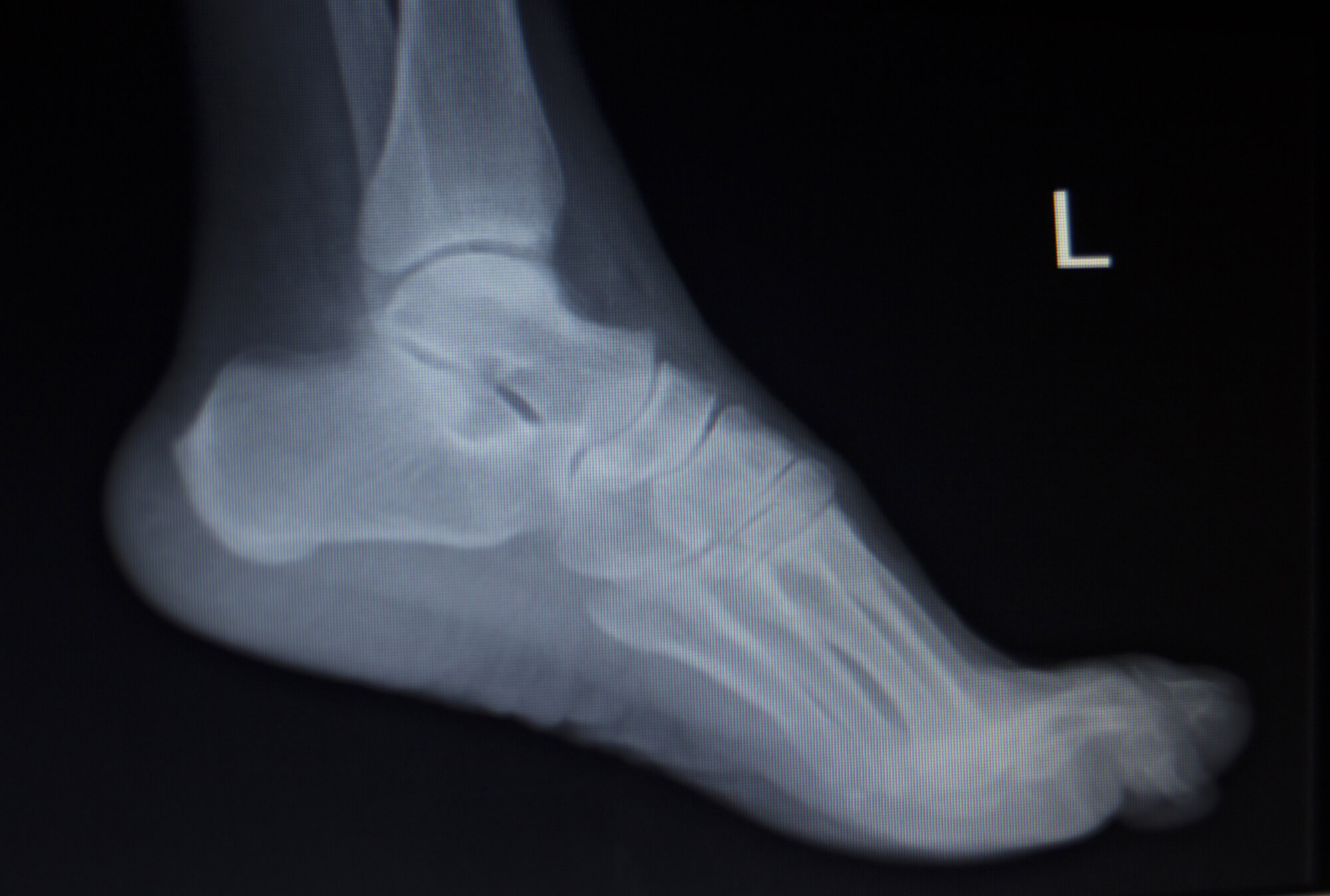
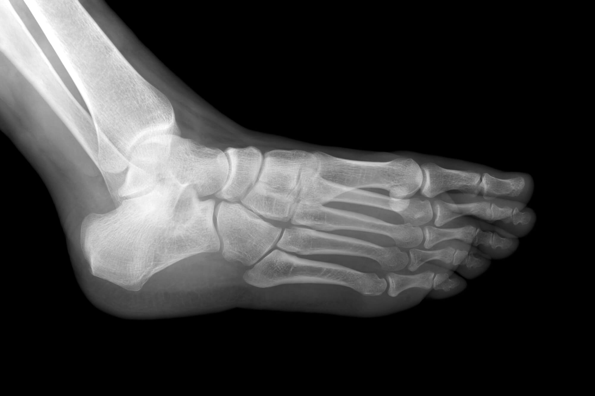
What Do Normal X-Ray Results Look Like?
Normal X-ray results will show the bones in the heel as solid white structures on the image. There should be no fractures or other abnormalities visible on the X-ray.
What Do Abnormal X-Ray Results Indicate?
Abnormal X-ray results may indicate the presence of a heel spur, a fracture, or other underlying condition that is causing the heel pain. The healthcare provider will interpret the X-ray results and discuss the findings with you, along with any recommended treatment options.
Treatment Options for Heel Pain Based on X-Ray Results
Treatment Options for Common Causes of Heel Pain
Treatment options for heel pain will depend on the underlying cause of the pain. Some common treatment options for conditions such as plantar fasciitis and heel spurs include:
- Stretching and strengthening exercises to improve flexibility and support the arch of the foot
- Orthotic inserts to help correct any foot abnormalities and provide support
- Ice and over-the-counter pain medication to reduce inflammation and pain
- Physical therapy to help improve mobility and reduce pain
Surgical Options for Heel Pain
In some cases, surgical intervention may be necessary to treat heel pain. This may include procedures such as heel spur removal or fracture repair. The healthcare provider will discuss the risks and benefits of surgery with you and help you decide if it is the right treatment option for your specific situation.
Conclusion
X-rays are a valuable diagnostic tool for understanding the cause of heel pain and developing a treatment plan. They are quick, non-invasive, and painless, and they can provide important information about the structure and condition of the bones and tissues in the heel. If you are experiencing heel pain, it is important to speak with a healthcare provider to determine the cause and find the most appropriate treatment option.

FAQ
What are X-rays and how do they work?
X-rays are a form of electromagnetic radiation with wavelengths shorter than visible light. They are used to create images of the inside of the body, including bones, organs, and tissues. X-rays work by passing through the body and being absorbed by different tissues at different rates. Dense tissues such as bones absorb more X-rays than softer tissues such as muscles and organs. The X-rays that pass through the body are detected by a special film or digital detector, which creates an image of the inside of the body.
Why are X-rays used for heel pain?
X-rays can be helpful in diagnosing the cause of heel pain because they allow the healthcare provider to see the structure and condition of the bones and tissues in the heel. For example, an X-ray can show if there is a heel spur present or if there is a fracture in the heel bone. It can also help to rule out other conditions that may be causing the pain, such as a stress fracture or a tumor.
What should I expect during an X-ray for heel pain?
X-rays for heel pain are usually performed on an outpatient basis, which means you will not need to stay in the hospital overnight. The procedure is typically quick and painless. You will be asked to stand on a platform with your heel positioned under the X-ray machine. The X-ray machine will emit a brief burst of X-rays that pass through your heel and are captured by the film or digital detector. You will be asked to hold still while the X-rays are being taken to ensure the best possible image quality. The X-ray process typically takes a few minutes.
How do I understand the results of an X-ray for heel pain?
Normal X-ray results will show the bones in the heel as solid white structures on the image. There should be no fractures or other abnormalities visible on the X-ray. Abnormal X-ray results may indicate the presence of a heel spur, a fracture, or other underlying condition that is causing the heel pain. The healthcare provider will interpret the X-ray results and discuss the findings with you, along with any recommended treatment options.
What treatment options are available for heel pain based on X-ray results?
Treatment options for heel pain will depend on the underlying cause of the pain. Some common treatment options for conditions such as plantar fasciitis and heel spurs include stretching and strengthening exercises, orthotic inserts, ice and over-the-counter pain medication, and physical therapy. In some cases, surgical intervention may be necessary to treat heel pain. This may include procedures such as heel spur removal or fracture repair. The healthcare provider will discuss the risks and benefits of surgery with you and help you decide if it is the right treatment option for your specific situation.